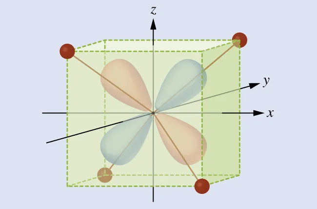
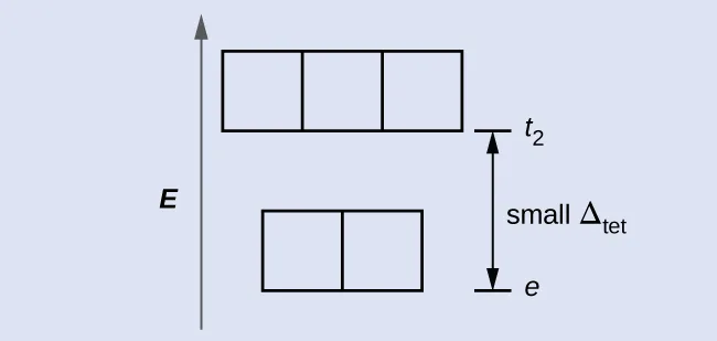
The behavior of coordination compounds cannot be adequately explained by the same theories used for main group element chemistry. The observed geometries of coordination complexes are not consistent with hybridized orbitals on the central metal overlapping with ligand orbitals, as would be predicted by valence bond theory. The observed colors indicate that the d orbitals often occur at different energy levels rather than all being degenerate, that is, of equal energy, as are the three p orbitals. To explain the stabilities, structures, colors, and magnetic properties of transition metal complexes, a different bonding model has been developed. Just as valence bond theory explains many aspects of bonding in main group chemistry, crystal field theory is useful in understanding and predicting the behavior of transition metal complexes.
Crystal Field Theory
To explain the observed behavior of transition metal complexes (such as how colors arise), a model involving electrostatic interactions between the electrons from the ligands and the electrons in the unhybridized d orbitals of the central metal atom has been developed. This electrostatic model is crystal field theory (CFT). It allows us to understand, interpret, and predict the colors, magnetic behavior, and some structures of coordination compounds of transition metals.
CFT focuses on the nonbonding electrons on the central metal ion in coordination complexes not on the metal-ligand bonds. Like valence bond theory, CFT tells only part of the story of the behavior of complexes. However, it tells the part that valence bond theory does not. In its pure form, CFT ignores any covalent bonding between ligands and metal ions. Both the ligand and the metal are treated as infinitesimally small point charges.
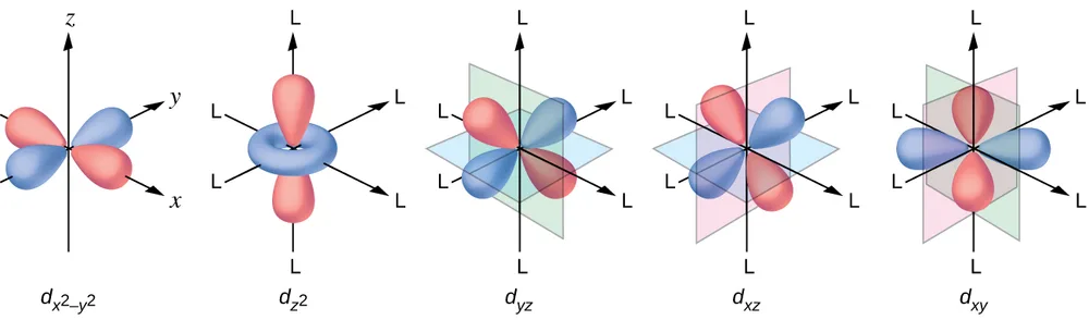
All electrons are negative, so the electrons donated from the ligands will repel the electrons of the central metal. Let us consider the behavior of the electrons in the unhybridized d orbitals in an octahedral complex. The five d orbitals consist of lobe-shaped regions and are arranged in space, as shown in Figure 19.33. In an octahedral complex, the six ligands coordinate along the axes.
In an uncomplexed metal ion in the gas phase, the electrons are distributed among the five d orbitals in accord with Hund's rule because the orbitals all have the same energy. However, when ligands coordinate to a metal ion, the energies of the d orbitals are no longer the same.
In octahedral complexes, the lobes in two of the five d orbitals, the and orbitals, point toward the ligands (Figure 19.33). These two orbitals are called the e orbitals (the symbol actually refers to the symmetry of the orbitals, but we will use it as a convenient name for these two orbitals in an octahedral complex). The other three orbitals, the , , and orbitals, have lobes that point between the ligands and are called the (again, the symbol really refers to the symmetry of the orbitals). As six ligands approach the metal ion along the axes of the octahedron, their point charges repel the electrons in the orbitals of the metal ion. However, the repulsions between the electrons in the orbitals (the and orbitals) and the ligands are greater than the repulsions between the electrons in the orbitals (the , , and orbitals) and the ligands. This is because the lobes of the orbitals point directly at the ligands, whereas the lobes of the orbitals point between them. Thus, electrons in the orbitals of the metal ion in an octahedral complex have higher potential energies than those of electrons in the orbitals. The difference in energy may be represented as shown in Figure 19.34.
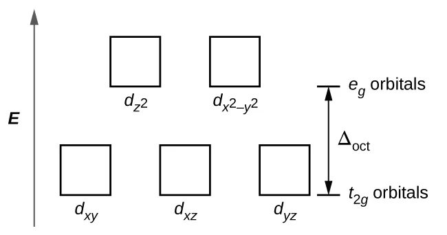
The difference in energy between the e and the t orbitals is called the crystal field splitting and is symbolized by Δ, where oct stands for octahedral.
The magnitude of Δ depends on many factors, including the nature of the six ligands located around the central metal ion, the charge on the metal, and whether the metal is using 3d, 4d, or 5d orbitals. Different ligands produce different crystal field splittings. The increasing crystal field splitting produced by ligands is expressed in the spectrochemical series, a short version of which is given here:
In this series, ligands on the left cause small crystal field splittings and are weak-field ligands, whereas those on the right cause larger splittings and are strong-field ligands. Thus, the Δ value for an octahedral complex with iodide ligands (I⁻) is much smaller than the Δ value for the same metal with cyanide ligands (CN⁻).
Electrons in the d orbitals follow the aufbau (“filling up”) principle, which says that the orbitals will be filled to give the lowest total energy, just as in main group chemistry. When two electrons occupy the same orbital, the like charges repel each other. The energy needed to pair up two electrons in a single orbital is called the pairing energy (P). Electrons will always singly occupy each orbital in a degenerate set before pairing. P is similar in magnitude to Δ. When electrons fill the d orbitals, the relative magnitudes of Δ and P determine which orbitals will be occupied.
In [Fe(CN)₆]⁴⁻, the strong field of six cyanide ligands produces a large Δ. Under these conditions, the electrons require less energy to pair than they require to be excited to the e orbitals (Δ > P). The six 3d electrons of the Fe²⁺ ion pair in the three t orbitals (Figure 19.35). Complexes in which the electrons are paired because of the large crystal field splitting are called because the number of unpaired electrons (spins) is minimized.
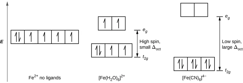
In [Fe(H₂O)₆]²⁺, on the other hand, the weak field of the water molecules produces only a small crystal field splitting (Δ < P). Because it requires less energy for the electrons to occupy the e orbitals than to pair together, there will be an electron in each of the five 3d orbitals before pairing occurs. For the six d electrons on the iron(II) center in [Fe(H₂O)₆]²⁺, there will be one pair of electrons and four unpaired electrons (Figure 19.35). Complexes such as the [Fe(H₂O)₆]²⁺ ion, in which the electrons are unpaired because the crystal field splitting is not large enough to cause them to pair, are called high-spin complexes because the number of unpaired electrons (spins) is maximized.
A similar line of reasoning shows why the [Fe(CN)₆]³⁻ ion is a low-spin complex with only one unpaired electron, whereas both the [Fe(H₂O)₆]³⁺ and [FeF₆]³⁻ ions are high-spin complexes with five unpaired electrons.
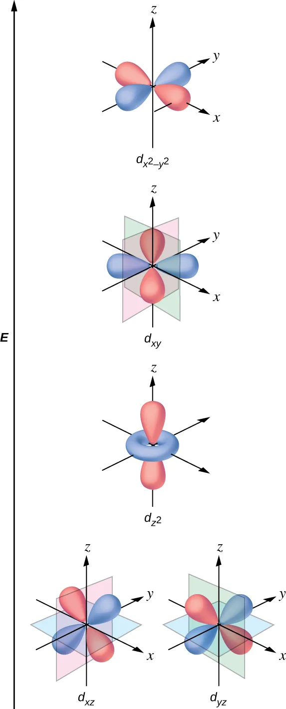
The other common geometry is square planar. It is possible to consider a square planar geometry as an octahedral structure with a pair of trans ligands removed. The removed ligands are assumed to be on the z-axis. This changes the distribution of the d orbitals, as orbitals on or near the z-axis become more stable, and those on or near the x- or y-axes become less stable. This results in the octahedral t and the e sets splitting and gives a more complicated pattern, as depicted below:
Magnetic Moments of Molecules and Ions
Experimental evidence of magnetic measurements supports the theory of high- and low-spin complexes. Remember that molecules such as O₂ that contain unpaired electrons are paramagnetic. Paramagnetic substances are attracted to magnetic fields. Many transition metal complexes have unpaired electrons and hence are paramagnetic. Molecules such as N₂ and ions such as Na⁺ and [Fe(CN)₆]⁴⁻ that contain no unpaired electrons are diamagnetic. Diamagnetic substances have a slight tendency to be repelled by magnetic fields.
When an electron in an atom or ion is unpaired, the magnetic moment due to its spin makes the entire atom or ion paramagnetic. The size of the magnetic moment of a system containing unpaired electrons is related directly to the number of such electrons: the greater the number of unpaired electrons, the larger the magnetic moment. Therefore, the observed magnetic moment is used to determine the number of unpaired electrons present. The measured magnetic moment of low-spin d⁶ [Fe(CN)₆]⁴⁻ confirms that iron is diamagnetic, whereas high-spin d⁶ [Fe(H₂O)₆]²⁺ has four unpaired electrons with a magnetic moment that confirms this arrangement.
Colors of Transition Metal Complexes
When atoms or molecules absorb light at the proper frequency, their electrons are excited to higher-energy orbitals. For many main group atoms and molecules, the absorbed photons are in the ultraviolet range of the electromagnetic spectrum, which cannot be detected by the human eye. For coordination compounds, the energy difference between the d orbitals often allows photons in the visible range to be absorbed.
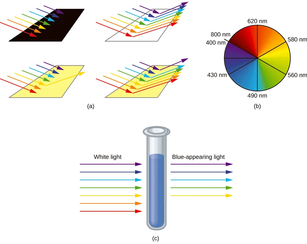
The human eye perceives a mixture of all the colors, in the proportions present in sunlight, as white light. Complementary colors, those located across from each other on a color wheel, are also used in color vision. The eye perceives a mixture of two complementary colors, in the proper proportions, as white light. Likewise, when a color is missing from white light, the eye sees its complement. For example, when red photons are absorbed from white light, the eyes see the color green. When violet photons are removed from white light, the eyes see lemon yellow. The blue color of the [Cu(NH₃)₄]²⁺ ion results because this ion absorbs orange and red light, leaving the complementary colors of blue and green (Figure 19.37).
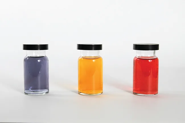
Small changes in the relative energies of the orbitals that electrons are transitioning between can lead to drastic shifts in the color of light absorbed. Therefore, the colors of coordination compounds depend on many factors. As shown in Figure 19.38, different aqueous metal ions can have different colors. In addition, different oxidation states of one metal can produce different colors, as shown for the vanadium complexes in the link below.
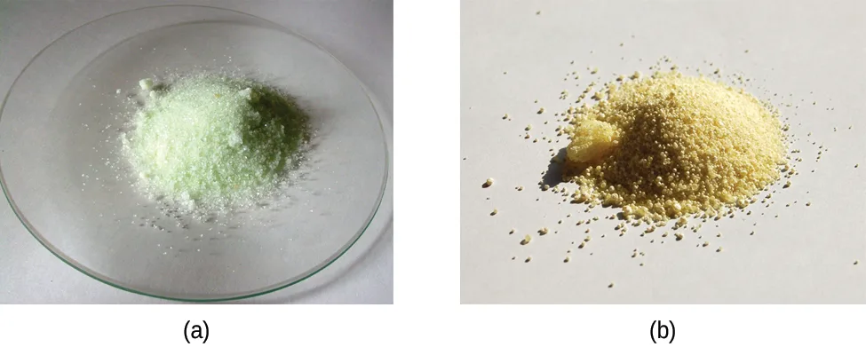
The specific ligands coordinated to the metal center also influence the color of coordination complexes. For example, the iron(II) complex [Fe(H₂O)₆]SO₄ appears blue-green because the high-spin complex absorbs photons in the red wavelengths (Figure 19.39). In contrast, the low-spin iron(II) complex K₄[Fe(CN)₆] appears pale yellow because it absorbs higher-energy violet photons.
In general, strong-field ligands cause a large split in the energies of d orbitals of the central metal atom (large Δ). Transition metal coordination compounds with these ligands are yellow, orange, or red because they absorb higher-energy violet or blue light. On the other hand, coordination compounds of transition metals with weak-field ligands are often blue-green, blue, or indigo because they absorb lower-energy yellow, orange, or red light.
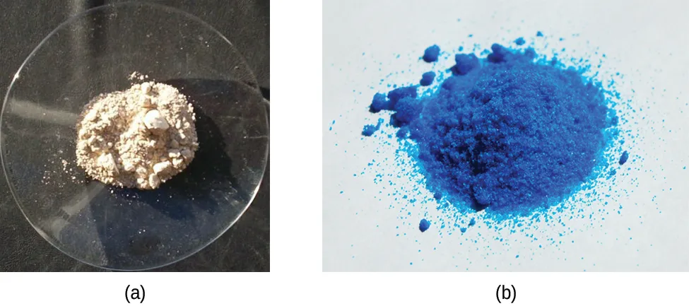
A coordination compound of the Cu⁺ ion has a d¹⁰ configuration, and all the e orbitals are filled. To excite an electron to a higher level, such as the 4p orbital, photons of very high energy are necessary. This energy corresponds to very short wavelengths in the ultraviolet region of the spectrum. No visible light is absorbed, so the eye sees no change, and the compound appears white or colorless. A solution containing [Cu(CN)₂]⁻, for example, is colorless. On the other hand, octahedral Cu²⁺ complexes have a vacancy in the e orbitals, and electrons can be excited to this level. The wavelength (energy) of the light absorbed corresponds to the visible part of the spectrum, and Cu²⁺ complexes are almost always colored—blue, blue-green violet, or yellow (Figure 19.40). Although CFT successfully describes many properties of coordination complexes, molecular orbital explanations (beyond the introductory scope provided here) are required to understand fully the behavior of coordination complexes.
Key Concepts and Summary
Crystal field theory treats interactions between the electrons on the metal and the ligands as a simple electrostatic effect. The presence of the ligands near the metal ion changes the energies of the metal d orbitals relative to their energies in the free ion. Both the color and the magnetic properties of a complex can be attributed to this crystal field splitting. The magnitude of the splitting (Δ) depends on the nature of the ligands bonded to the metal. Strong-field ligands produce large splitting and favor low-spin complexes, in which the t orbitals are completely filled before any electrons occupy the e orbitals. Weak-field ligands favor formation of high-spin complexes. The and the orbitals are singly occupied before any are doubly occupied.